Before an antitumor drug enters preclinical development, the first question to answer is not “does it work?” but “in which tumor type does it work best?”
For the same molecule, tumor growth inhibition can reach 80% in Tumor Type A, yet drop to merely 30% in Tumor Type B. Selecting an inappropriate CDX model distorts data quality and yields a false‑positive efficacy conclusion instead of a true negative result, which not only wastes R&D resources but also reduces the success rate of subsequent clinical trials.
This is why CDX (Cell‑Line‑Derived Xenograft) models remain the workhorse for antitumor pharmacodynamic assessment. Within minimal time and at low cost, they tell you which indication is worth further investment for your molecule.
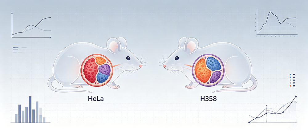
Let’s look at data showing two distinct performances of Test Article A1.
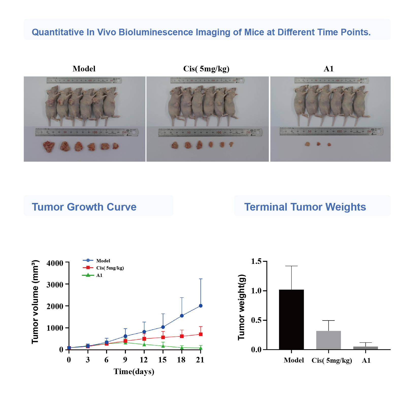
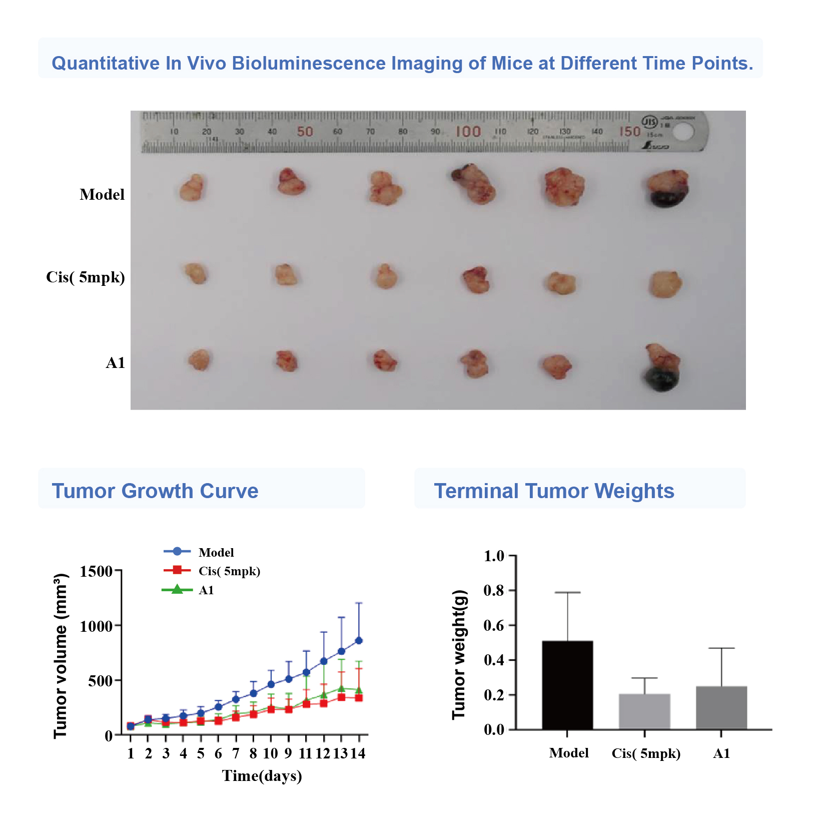
We established two subcutaneous CDX models using Balb/c‑nu mice to validate the same test article A1.
Case 1: HeLa Cervical Cancer CDX Model
Test article A1 exhibits potent antitumor activity, superior to cisplatin.
Case 2: H358 Lung Cancer CDX Model
Test article A1 shows antitumor efficacy comparable to cisplatin.
Valuable insights emerge when comparing both datasets side‑by‑side.
| Metrics | HeLa Cervical Cancer | H358 Lung Cancer |
| Absolute activity of A1 | Potent (near‑complete tumor inhibition) | Moderate (effective without full tumor ablation) |
| A1 vs. Cisplatin | Markedly superior | Roughly equivalent |
| Decision‑making value | High‑priority indication for progression | Further data required for decision‑making |
If only the HeLa data were considered, A1’s superior efficacy could be over‑extrapolated. If only the H358 dataset is reviewed, A1 might be dismissed as “no better than existing therapies”, and its differentiated value for cervical cancer would be overlooked.
Divergent performances of one molecule across different tumor types are not an exception — they are common. Only with properly selected CDX models can preclinical data support sound decision‑making, enabling robust indication positioning and higher success rates for clinical trials. The core value lies not in answering “is this drug effective?”, but precisely identifying “which indication deserves prioritized advancement”.
Why Subcutaneous CDX Remains the Go‑To Starting Point
Subcutaneous implantation offers the most straightforward readout and highest throughput among CDX approaches. Tumor volume can be measured non‑invasively with vernier calipers without imaging equipment. Guided by the “same drug, different tumors” screening logic, it is irreplaceable for the following scenarios:
- Early‑stage screening: Rapid in‑vivo ranking of multiple candidate molecules across tumor types with well‑controlled cost and timeline.
- Dose‑finding studies: High‑frequency tumor volume measurement (2‑3 times weekly) to finely characterize dose‑response relationships.
- Positive control calibration: Deploy standard agents such as cisplatin to define model response windows and benchmark the relative potency of test articles.
- Combination therapy screening: Preliminary assessment of combinatorial regimens; promising signals can then be validated in orthotopic or PDX models.
Note: Subcutaneous CDX lacks native tumor orthotopic microenvironments. Immunodeficient mice are not suitable for immunotherapy evaluation. For projects demanding higher clinical relevance, consider advancing to orthotopic CDX or PDX models (see comparison table in appendix).
Jennio CDX Platform: Model Diversity Determines Screening Depth
The value of a model lies half in the model itself and half in the diversity of available cell‑line options. Without a diverse panel of cell lines, comparisons under the “same drug, different tumors” paradigm cannot be performed.
Jennio CDX cell bank covers 13 cancer types with nearly 400 cell lines (partial list shown below):
| Cancer Type | Number of Cell Lines |
| Lung Cancer | 81 |
| Lymphoma | 45 |
| Liver Cancer | 38 |
| Esophageal Cancer | 23 |
| Thyroid Cancer | 19 |
| Bladder Cancer | 15 |
| Gallbladder Cancer | 8 |
| Leukemia | 50 |
| Colorectal Cancer | 42 |
| Bone / Soft Tissue Sarcoma | 25 |
| Cervical / Endometrial Cancer | 21 |
| Nasopharyngeal Carcinoma | 15 |
| Pancreatic Cancer | 15 |
| (Partial list only) |
We maintain a standardized tumor‑model portfolio. Our CDX service supports subcutaneous, orthotopic and intravenous implantation using human‑derived or murine cell lines. PDX (Patient‑Derived Xenograft) models are also available to match your project stage.
A broader selection of cell lines in the cell bank helps you find the “optimal indication” for candidate A1 in the early project phase, instead of conducting repeated validation on unsuitable tumor types.
Model Progression Path: Subcutaneous CDX as a Starting Point, Not the Endpoint
| Model | Immune Status | Tumor Microenvironment | Timeline / Cost | Application Stage |
| Subcutaneous CDX | Immunodeficient | No native orthotopic microenvironment | Short / Low | Early‑phase screening, tumor‑type prioritization, dose‑finding |
| Orthotopic CDX | Immunodeficient | Native orthotopic microenvironment | Moderate / Moderate | Efficacy confirmation, metastasis and microenvironment research |
| PDX | Immunodeficient | Partially preserved human tumor features | Long / High | Clinical‑relevance validation, precision‑medicine studies |
| Syngeneic Transplantation | Fully intact immune system | Native microenvironment | Moderate / Moderate | Immunotherapy assessment |
Where should you go next after identifying positive hits from subcutaneous CDX screening?
In our next article, we will use gastric, liver and cervical orthotopic tumor models as examples. We will walk through complete datasets combining dynamic monitoring via in‑vivo bioluminescence imaging paired with terminal tumor‑weight readouts, illustrating the workflow for efficacy evaluation advancing from subcutaneous to orthotopic models. Stay tuned!
Appendix: Four Key Questions before Selecting Tumor Types
You do not need broad screening for your first round. Answer these four questions to shortlist 2‑3 candidate tumor types:
- Where is your target highly expressed? (Refer to TCGA / CCLE datasets; prioritize cancer types with target expression within the top 30%).
- Does this tumor type depend on your target pathway? (High target expression does not guarantee pathway‑dependence; cross‑reference published literature.)
- For drugs targeting the same target, in which tumor types have positive efficacy data been reported? (Tumor‑type distribution of marketed or clinical‑stage drugs provides valuable prior probability.)
- Is there an established standard‑of‑treatment agent usable as positive control for this tumor type? (Without reference benchmarks, you cannot draw conclusions on “superiority” or “equivalence”).
After narrowing down candidates with these four questions, run parallel subcutaneous CDX assays. Select the combinations showing strongest superior efficacy signals, then allocate resources for further advancement.
Data Source Note
- All pharmacodynamic data in this article are generated from in‑house efficacy studies conducted by Jennio Biotech
- Cancer‑type coverage and cell‑line counts for the CDX cell bank originate from Jennio internal platform records (data as of publication; subject to real‑time inventory updates).