Tumor In Situ Model

Tumor In Situ Model

CDX models play a critical role in the preclinical evaluation of anti-tumor drugs. The establishment of tumor animal models provides powerful tools for studying the mechanisms of tumor development and metastasis, as well as for screening and evaluating the efficacy of anti-tumor drugs. Currently, the available rodent tumor models include spontaneous tumor models, induced tumor models, genetically engineered mouse tumor models, as well as xenograft/allograft tumor models. Subcutaneous (s.c.) grafts, one of the transplantation models, are more widely applicable for preclinical research.

Details

Overcoming the Bottleneck of Clinical Translation in Anticancer Drug Development — High-Predictivity Orthotopic Models Empower Precise Drug Efficacy Evaluation

  • Industry Pain Point: Traditional subcutaneous xenograft models lack the organ microenvironment, resulting in a disconnect between preclinical data and human response.
  • Core Value: Based on orthotopic transplantation technology to restore tumor-host interaction, we significantly improve the accuracy of drug efficacy prediction and accelerate the clinical translation of novel drugs.

1、Service Overview

  • Orthotopic tumor models are a critical component of the preclinical anticancer drug evaluation system.
  • Traditional subcutaneous xenografts lack an organ-specific microenvironment, making it difficult to accurately simulate drug distribution, metabolism, and therapeutic response in humans. This contributes to the clinical failure of approximately 70% of drug candidates.
  • Jennio Bio establishes high-fidelity orthotopic tumor models (including liver cancer, cervical cancer, gastric cancer, etc.) by implanting tumor cells or tissue fragments into the corresponding organs of immunodeficient mice (e.g., liver capsule, cervix, gastric wall). This approach precisely recapitulates the human tumor growth microenvironment, providing pharmaceutical companies with:
    • More reliable efficacy data: Orthotopic models preserve key features such as tumor angiogenesis, invasion, and metastasis, thus enabling clinical-relevant drug responses.
    • More precise mechanistic studies: Supports mechanism validation for targeted therapies and immunotherapies in a physiologically relevant microenvironment.
    • Reduced clinical failure risk: Collaborations over the past five years demonstrate a significant improvement in clinical success rates for drugs advanced based on orthotopic model data.

2、Our Services

Application scenarios segmented by tumor type and R&D stage:

  • Orthotopic Models for High-Relapse Cancers
    • Orthotopic Liver Cancer Model: Intra liver implantation of SMMC7721 luc cells, with real time tumor growth monitoring via in vivo imaging. (Case: Lenvatinib markedly suppressed fluorescence signals and reduced tumor weight by approximately 60%.)
    • Orthotopic Cervical Cancer Model: Cervical transplantation of HeLa luc cells. Tumor volume in the uterine cavity was reduced by approximately 50% in the cisplatin treatment group.
    • Orthotopic Gastric Cancer Model: Gastric wall implantation of MKN45 luc cells. The oxaliplatin group showed reduced fluorescence signals and significantly decreased tumor weight.
  • Metastatic Tumor Models
    • Lung Metastasis Model: Nalm 6 leukemia cells are injected via the tail vein. The CAR T treatment group shows a significant reduction in fluorescence signals.
    • Bone Metastasis Model: Breast cancer cells are injected intra-tibially to evaluate the inhibitory effect of targeted agents on bone destruction.
  • Immunotherapy Evaluation Models
    • Humanized Immune System Orthotopic Model: Orthotopic tumors are established after implanting human PBMC or CD34+ stem cells into NCG mice, to evaluate the efficacy of PD-1/CTLA-4 antibodies, and detect tumor-infiltrating lymphocytes (TIL) and cytokine secretion.
  • Technical Approaches:
    • In vivo imaging (fluorescence / bioluminescence)
    • Histopathology (H&E / IHC)
    • Flow cytometry (immune cell subtyping in the microenvironment)
    • Molecular detection (qPCR / ELISA)

3、Technology Platforms and Advantages

  • Core Platforms
    • PDX Orthotopic Transplantation Platform: Direct transplantation of patient tumor tissue into the corresponding organs of mice (e.g., liver cancer implanted into the liver capsule), preserving tumor heterogeneity and clinical relevance.
    • Orthotopic-Metastasis Integrated Model: Simultaneous evaluation of drug efficacy in primary and metastatic lesions in the same animal (e.g., orthotopic prostate cancer + bone metastasis model).
  • Facilities & Qualifications
    • SPF Animal Center: 5,000-cage capacity, equipped with in vivo imaging systems, small animal ultrasound, and other instruments.
    • GLP-aligned Standards: Experimental procedures comply with GLP specifications (SOP management, dual independent data review) to ensure data traceability.
  • Differentiation Advantages
    • Exclusive Chinese Primary Tumor Bank: Over 300 native tumor cell lines, providing a better fit for drug efficacy responses in the Asian population.
    • Orthotopic Model Success Rate >85%: The technical team has 10 years of operational experience, with transplantation survival rates leading the industry.

4、Service Process

  • Requirement Confirmation (1–3 days): Define tumor type, cell line/PDX source, dosing regimen, and endpoint indicators.
  • Model Establishment (2–4 weeks): Orthotopic transplantation + model validation (imaging/histology).
  • Efficacy Study (3–8 weeks): Grouped administration; regular monitoring of tumor volume (in vivo imaging), animal body weight, and survival rate.
  • Data Analysis (2 weeks): Provide raw imaging data, tumor weight statistics, immunohistochemistry images, and statistical analysis report.
  • Deliverables: Final GLP-aligned report (bilingual Chinese & English), including experimental procedures, raw data, and result interpretation.

5、Quality Control and Compliance
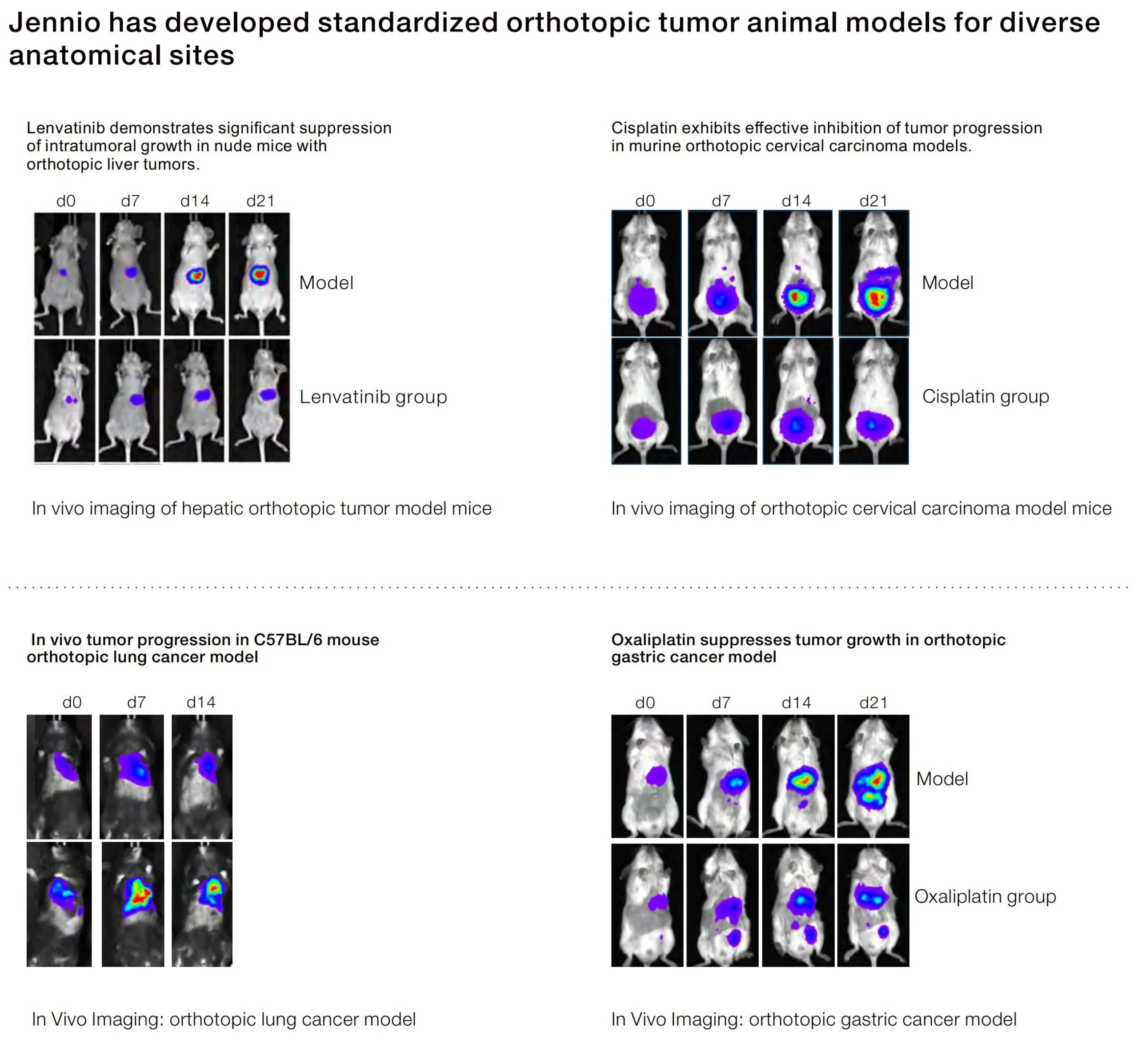
  • Compliance Standards: Laboratory animal welfare complies with AAALAC guidelines, and data management adheres to electronic record regulations.
  • Quality Control Nodes:
    • Pre-transplantation: STR identification of cells/tissues, pathogen detection.
    • During experiment: Double-blind analysis of imaging data, cross-review of tissue sections.
    • Before delivery: Three-level review (Technician → Project Manager → Quality Control Department).

6、Case Studies

Case 1: Orthotopic Gastric Cancer Model Efficacy Evaluation

  • Customer Requirement: To evaluate the inhibitory effect of oxaliplatin on orthotopic growth of gastric cancer.
  • Protocol: MKN45 luc cells were implanted into the gastric wall of NCG mice, followed by 3 weeks of grouped drug administration.
  • Results: Fluorescence signal in the treatment group was reduced by approximately 70% (p<0.01), and tumor weight decreased by about 60%.
  • Figures: Tumor growth curve (fluorescence intensity over time), bar chart for tumor weight comparison.

Case 2: CAR-T in Leukemia Lung Metastasis Model

  • Results: Tumor signal in the CAR T group was reduced by 70% compared with the control group.

For more cases, please refer to the special page of Jennio PDX Efficacy Platform.

7、Collaboration Advantages

  • Full-cycle Support: From model customization to IND-enabling report preparation, covering drug discovery to preclinical research.
  • High Efficiency: Standard project duration shortened by 30% vs. industry average; urgent projects initiated within 72 hours.
  • Global Collaboration Experience: Served clients including Ribo Pharma, Hengrui Medicine, and supports multi-center data mutual recognition.

8、FAQ

  • Q: Do you support orthotopic models in non-human primates?

A: Not currently available, but we provide cross-species pharmacokinetic and toxicokinetic study designs in mice and cynomolgus monkeys.

  • Q: Can customer-owned cell lines be used for model establishment?

A: Yes, we accept client-provided cell lines, with submission of cell STR authentication reports and culture records required.

  • Q:Can the data be used for US-China dual filings?

A: All studies are performed in accordance with ICH guidelines, and report formats meet FDA/EMA requirements.

9、Laboratory Highlights

  • Image 1: SPF Animal Facility (5,000 cage capacity, individually ventilated cages)

Note: Ensures pathogen-free model animals, improving data reliability by 40%.

  • Image 2: Small Animal In Vivo Imaging System (IVIS Spectrum)

Note: Real-time quantitative monitoring of orthotopic tumor growth with high specificity and precision.

  • Image 3: Microsurgical Operating Station (for tumor orthotopic transplantation)

Note: Supports high-precision intra-organ injection with a transplantation success rate >85%.


10、Contact Us

  • Contact Us Now:
    • Tel: +86 18802035152
    • Email: 3691125803@qq.com
  • Resource Download:
    • White Paper on Orthotopic Tumor Model Technology
    • PDX Efficacy Case Collection
  • Free Customized Protocol:

Submit your requirements and get an exclusive experimental design within 48 hours.


Listen to the voice of every customer

Share details of your inquiry, and we’ll contact you shortly.​

Contact us

Room 238, Building A, Guangzheng Science and Technology Park, No. 11 Nanyunwu Road, Huangpu District, Guangzhou, Guangdong Province, China

Business Cooperation

3691125803@qq.com

Mobile phone number/WeChat ID

+86 18802035152