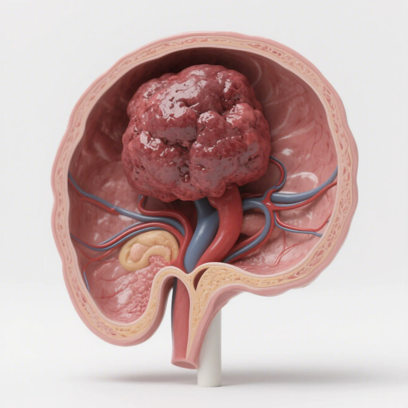
We choose between orthotopic and subcutaneous tumor models as required by the balance between throughput and organ-specific biology. We compare orthotopic and subcutaneous tumor models against the research question. Across the model comparison, he key decision criteria and remaining evidence gaps are documented before study execution begins.
The evidence boundary remains explicit in this orthotopic and subcutaneous model comparison program. We do not freeze the interpretation; relevant follow-up data can modify it. Subcutaneous grafts are easier to establish and measure with calipers, making them practical for screening, dose finding, and serial response tracking.
Our planning discussion therefore covers both the biological hypothesis and the operational path used to test it. As the tumor-model comparison progresses, we adjust the evidence package according to the importance and potential consequences of the development decision. Uncontrolled variability may obscure a true effect or make ordinary noise look meaningful.
Orthotopic grafts place the tumor in the relevant organ, better representing the local microenvironment, angiogenesis, invasion, and metastasis. We consider the tissue-specific drug distribution and tumor behavior captured by orthotopic models, together with their greater technical and monitoring requirements, when interpreting the results. We support both approaches and add PDX, metastatic, and humanized options.
A staged strategy may use subcutaneous models to prioritize candidates and orthotopic models for confirmation. Our discussion moves from model fit to record quality and then to action. Documenting the reasoning now reduces ambiguity during later scientific evaluation.

Why Subcutaneous Models Remain Valuable
The first control step is conceptual. The study team examines whether the experiment can answer the defined development concern. Neither format is universally superior; model validity, controls, monitoring, pathology confirmation, statistics, welfare, and interpretation determine usefulness.
In practical execution, we explain that conventional subcutaneous grafts are convenient and widely used, but they do not reproduce the organ-specific microenvironment that influences tumor-host interaction. Our evidence package contains angiogenesis, invasion, metastasis, drug distribution, metabolism, and treatment response as relevant to the study objective.
During tumor-model comparison planning, a tumor-model comparison design starts from a defined experimental unit. Our tumor-model comparison plan defines the starting material, initial state, and control in one connected rationale. In this review, predefined study-initiation criteria provide a consistent starting point for the tumor-model comparison
For orthotopic model establishment, our orthotopic or in situ platform implants tumor cells or patient-derived tissue into the anatomically corresponding organ of immunodeficient mice, including liver, stomach, cervix, and other relevant sites. Our comparison records which biological question each tumor model can answer and which it cannot.
In an orthotopic vs subcutaneous tumor model comparison, we focus on the evidence each format can produce. During active tumor-model comparison execution, our applications include orthotopic liver, gastric, and cervical cancer, lung and bone metastasis.
Related tumor-model comparison work can cover integrated primary-and-metastatic models, PDX orthotopic transplantation, and humanized immune-system models for immunotherapy. Biological system selection is an argument about relevance, not familiarity. At the confirmation stage, we align the tumor-model comparison system with candidate biology and development stage.
What Orthotopic Models Add to the Evidence
Across the model comparison, the evidence becomes stronger when independent observations converge. Depending on the selected tumor models and study objectives, technical readouts include fluorescence or bioluminescence imaging, small-animal ultrasound, H&E. Additional readouts include IHC, flow-cytometric immune profiling, qPCR, ELISA, body weight, survival, tumor weight, and pathology.
Using distinct measurement methods prevents accessibility of the tumor from biasing the biological conclusion. Our process includes requirement confirmation, cell-line or PDX-source selection, endpoint and dosing alignment, and tumor-model establishment and validation, which typically require two to four weeks depending on the selected model and study design.
The study team examines measurement reliability before attributing a difference to model location. For tumor-model comparison, we investigate failed runs alongside controls and signal stability. Our orthotopic vs subcutaneous tumor model strategy treats screening and confirmation as complementary stages. Controlled execution turns the design into usable supporting information.
We connect time course, checks, instruments, and raw observations in one record. Our schedule includes three to eight weeks of efficacy work followed by data analysis with raw images, statistics, IHC images, and a GLP-aligned report.
Our model comparisons at Jennio Biotech retain the partner’s decision criteria throughout review. We compare growth, exposure, pathology, invasion, and metastasis guided by the purpose of the study. External measurements and organ-specific tissue findings reveal different aspects of treatment response.
This restraint prevents the tumor-model comparison finding from being carried beyond its evidentiary reach. During study delivery, the defined evidence boundary makes the advantages and limitations of alternative model systems easier to assess.
Use a Staged Strategy Instead of a Binary Choice
We let the tumor-model comparison conclusion determine whether the program moves, adapts, or seeks confirmation. Each post-investigation route is tied to a finding identified before the record is closed. Quality controls include STR authentication, pathogen testing, SOP management, double-blind imaging check, cross-review of tissue sections, electronic records, and multilevel review before delivery.
For the connected decision, our operations maintain an SPF animal center with in vivo imaging, small-animal ultrasound, and microsurgical support. The tumor-model comparison evidence becomes more credible when uncertainty remains visible. System, dose, timing, and measurement define the scope of our tumor-model comparison interpretation. The program can take its next tumor-model comparison step when it becomes defensible through the package.
In practice, the most appropriate model is determined by the study question: a subcutaneous model can be preferable for efficient screening and direct caliper measurement. The review explains that an orthotopic model is more relevant when local growth, organ physiology, invasion, metastasis, or tissue-specific exposure is central.
The interpretation states what remains uncertain and which limitations are inherent to the selected model system.. Neither format removes the need for appropriate controls, validated implantation, prospective success criteria, serial monitoring, ethical oversight, and careful interpretation of disease model limitations.
We define how tumor-model comparison will close, transfer, and support later decisions. Our final tumor-model comparison record is structured for audit, transfer, or a complementary follow-up study. Our Jennio Biotech report states which tumor model fits the question and where the alternative may be more suitable.