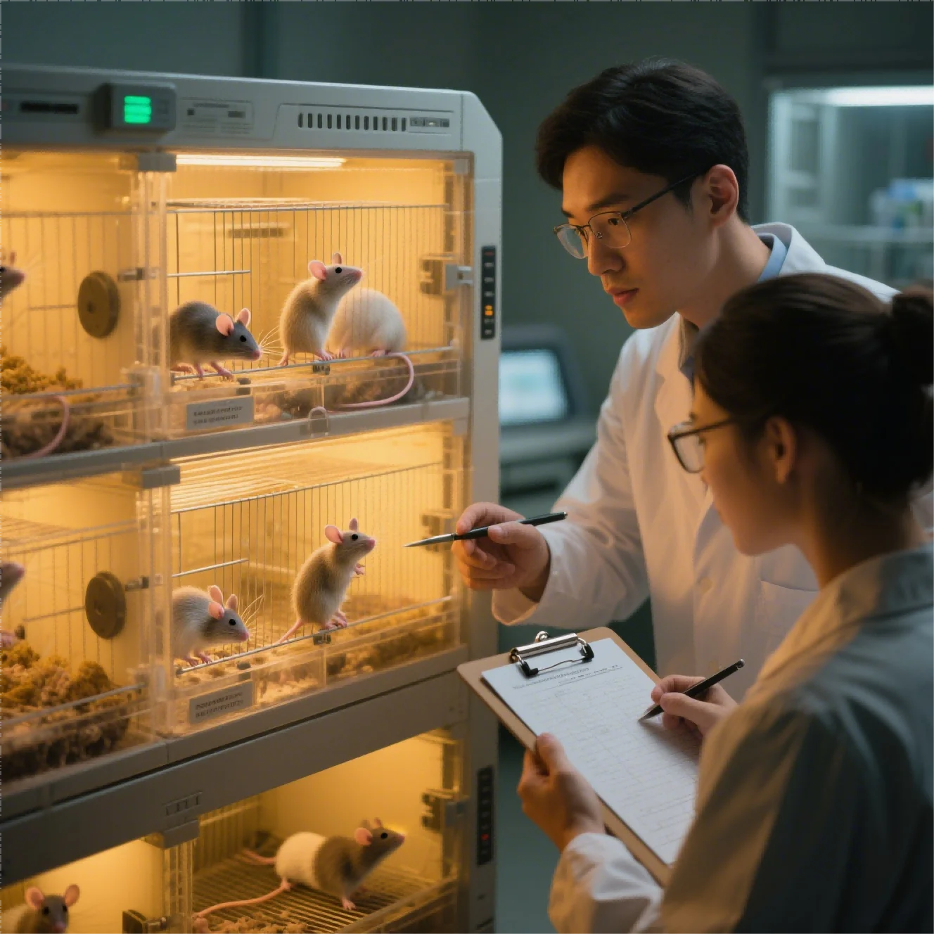
The study team uses the term in situ tumor model for preclinical systems that place tumor material in its anatomically relevant location, not for work performed in patients. We keep the intended biological question and model limitations explicit throughout in situ tumor study planning.
The in-situ tumor study plan begins with the decision gap that must be reduced. A predefined decision framework and documentation trail clarify potential next steps before the final report is written. Our orthotopic PDX approach transfers patient tumor tissue into the corresponding organ of a mouse to retain selected histological and molecular features of the original tumor and enhance translational relevance
The study team uses in situ model system patient tumor terminology only for patient-derived material placed in an anatomically relevant animal model. The function is to study tumor growth, invasion, metastasis, therapeutic response, and biomarkers in an organ-specific environment before clinical testing.
A fragmented workflow also makes it difficult to reconcile cellular activity, exposure, pathology, and safety observations. We deliver anatomical placement, imaging, pathology, and source records so the in situ finding can be independently reviewed.
The goal is a defensible determination, supported by records that another scientific team can review. It can support efficacy comparisons and translational hypotheses but cannot reproduce every aspect of a patient’s immune system or clinical course.
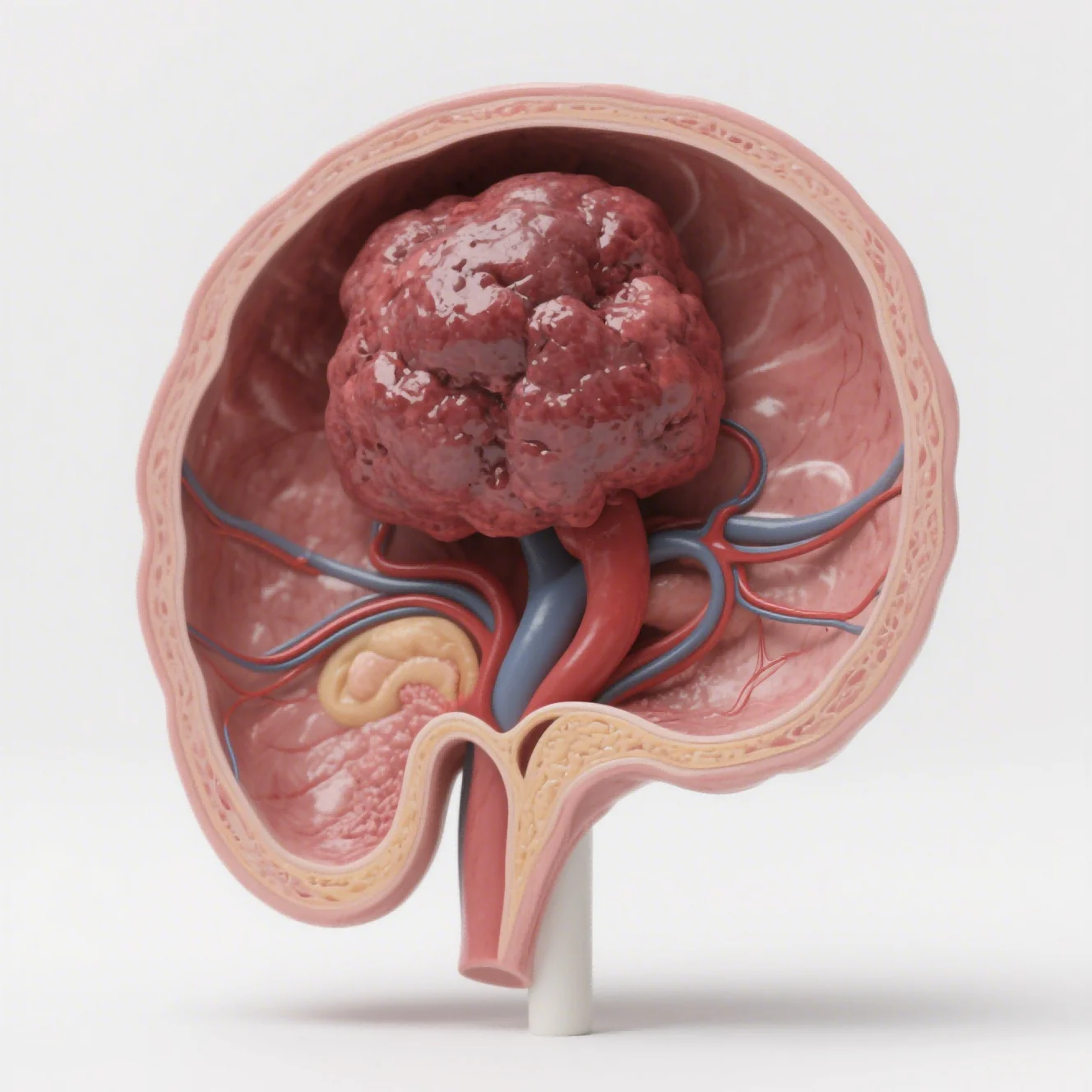
What Patient-Derived Material Preserves
Accurate wording needs to distinguish patient-derived material, patient-relevant biology, and actual patient research to avoid an ethically and scientifically misleading claim. Before discussing throughput, we establish relevance. We judge the model’s value within the research plan by its relevance to the target biology and mechanism of action..
A familiar workflow cannot substitute for a relevant in situ tumor study interpretation. Downstream analysis cannot compensate for biological relevance that is absent from the selected starting material or model design. We explain that conventional subcutaneous grafts are convenient and widely used, but they do not reproduce the organ-specific microenvironment that influences tumor-host interaction.
The delivered record brings together angiogenesis, invasion, metastasis, drug distribution, metabolism, and treatment response. Our material review for in situ tumor study covers origin, identity, quality, and intended use. Our orthotopic or in situ platform implants tumor cells or patient-derived tumor tissue into the anatomically corresponding organ of immunodeficient mice, including liver, stomach, cervix, and other sites.
An approach is informative only within the biology it represents. We specify that boundary before treating a readout as supporting information. Through Jennio Biotech, we connect every in-situ tumor-study choice to its stated purpose. We evaluate in situ tumor study capability through qualification evidence, not through service naming.
We review the evidence that qualifies the selected model or assay. For this in situ tumor study question, a clearly defined baseline and comparator make subsequent model comparisons more interpretable. We make the in-situ tumor-study conclusion traceable from source evidence to interpretation for partner review.
How an Organ-Specific System Supports Translation
Our applications include orthotopic models of liver, gastric, and cervical cancers; experimental models of lung and bone metastasis; and study designs that evaluate primary and metastatic tumor progression.. The requested decision may require us to include PDX orthotopic transplantation, and humanized immune-system study models for immunotherapy. During delivery, small inconsistencies deserve early research discussion.
This safeguard reduces in situ tumor study rework and protects the intended comparison. Our reading of in situ tumor study is governed by mechanism and data, not ease of execution. We choose measurements to fit the explanation required from the research plan. We combine response, mechanism, exposure, or pathology where the research concern requires it.
Technical readouts include fluorescence or bioluminescence imaging, small-animal ultrasound, H&E, IHC, flow-cytometric immune profiling, qPCR, ELISA, body weight, survival, tumor weight, and pathology. Our process includes requirement confirmation, cell-line or PDX-source selection, endpoint and dosing alignment, two to four weeks of experimental system construction and validation.
Operational consistency protects technical interpretation. Instrument status and sampling time are controlled together within the in-situ tumor study. The in-situ model system patient tumor work connects organ-specific behavior with a carefully bounded translational question. The in-situ tumor study report reflects quality controls applied while the work was performed.
The evidence package additionally includes three to eight weeks of efficacy work, and subsequent data analysis with raw images, statistics, IHC images, and a structured study report prepared under applicable quality-control procedures. We trace in situ tumor study findings to the procedure, biological input, and completed review.
Interpret Patient Relevance Without Overstatement
Quality controls include STR authentication, pathogen testing, SOP management, double-blind imaging audit, cross-review of tissue sections, electronic records, and multilevel review before delivery. Our operations maintain an SPF animal center with in vivo imaging, small-animal ultrasound, and microsurgical support.
The final stage turns observations into a bounded development next-step call. The investigation closeout keeps confirmed evidence separate from unresolved work. The in-situ study closes with an explicit account of model relevance, technical limits, observed response, and unanswered questions. We relate every claim to predefined criteria and available raw supporting information.
The study team chooses the system by the study question: subcutaneous systems support efficient screening and caliper measurement. Orthotopic systems also address local growth, organ physiology, invasion, metastasis, and tissue-specific exposure. Determination quality improves when evidence and uncertainty travel together.
Our in-situ tumor study report prevents an unresolved issue from being absorbed into the main claim. The Jennio Biotech closeout keeps anatomical relevance, technical difficulty, and treatment response in view. We use This point to test the package’s completeness from another team’s perspective.
At handoff, the in-situ tumor study data remain connected to method, interpretation, and unresolved questions. Neither format removes the need for appropriate controls, validated implantation, prospective success criteria, serial monitoring, ethical oversight, and careful interpretation of biological model limitations. This distinction protects scientific accuracy while keeping the ethical boundary explicit.