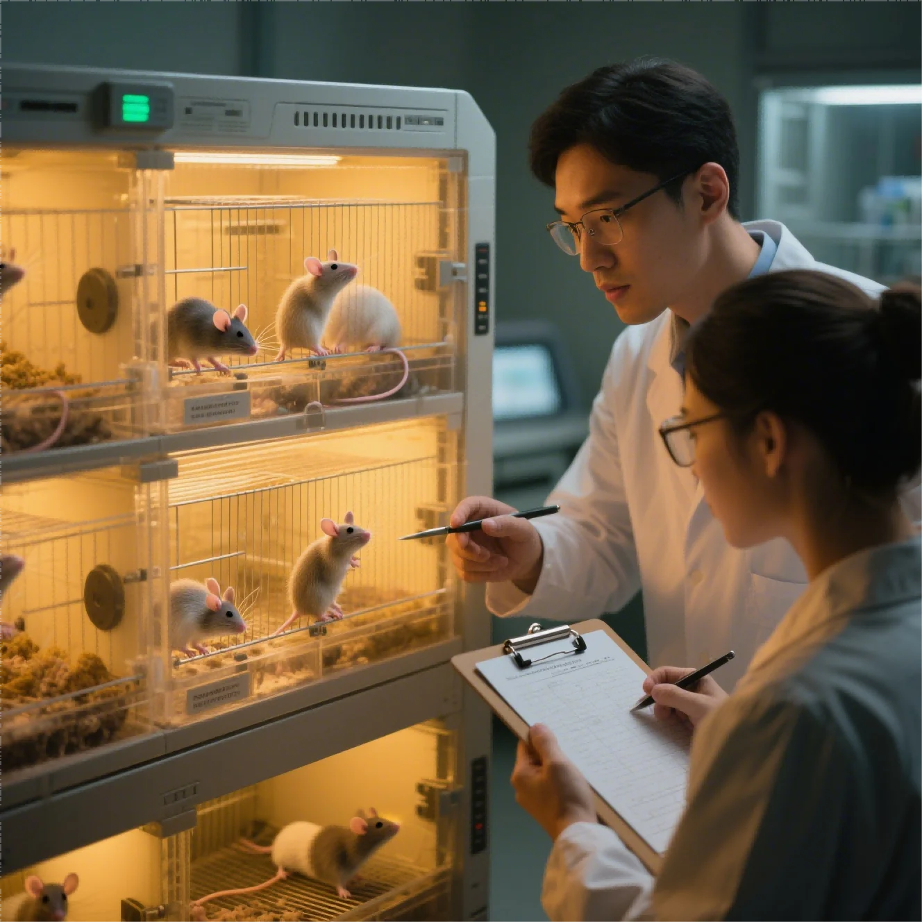
We place tumor cells or tissue in the pancreas when an orthotopic pancreatic tumor model is needed to study organ-relevant growth and spread. The study team retains the source data and experimental records needed to assess how vasculature, invasion routes, metastasis, and organ-specific drug exposure influence the result.
The study team traces each orthotopic pancreatic study result back to the materials, procedures, controls checks, and raw observations that support it. The study team traces the result back to its biological material, method, checks, and raw observations. A useful plan distinguishes a promising signal from data that is sufficiently controlled for the next step.
Within this development effort, a later system can be evaluated by whether it preserves the required orthotopic pancreatic study function. A pancreatic model would require skilled microsurgery and a well-characterized pancreatic tumor source. We interpret the main result alongside imaging or another method for monitoring a non-palpable lesion, humane endpoints, and pathology confirming tumor location and spread.
Convenient endpoints can create false confidence when they do not reflect the candidate’s mechanism or intended route. This approach keeps the development question in view while allowing the strength and limitations of the model to be assessed from relevant supporting data
The study needs to define implantation success, baseline burden, randomization, dosing, survival or tumor endpoints, metastatic assessment, and tissue biomarkers. We use an orthotopic pancreatic tumor model when organ-specific growth and spread are central to the question.
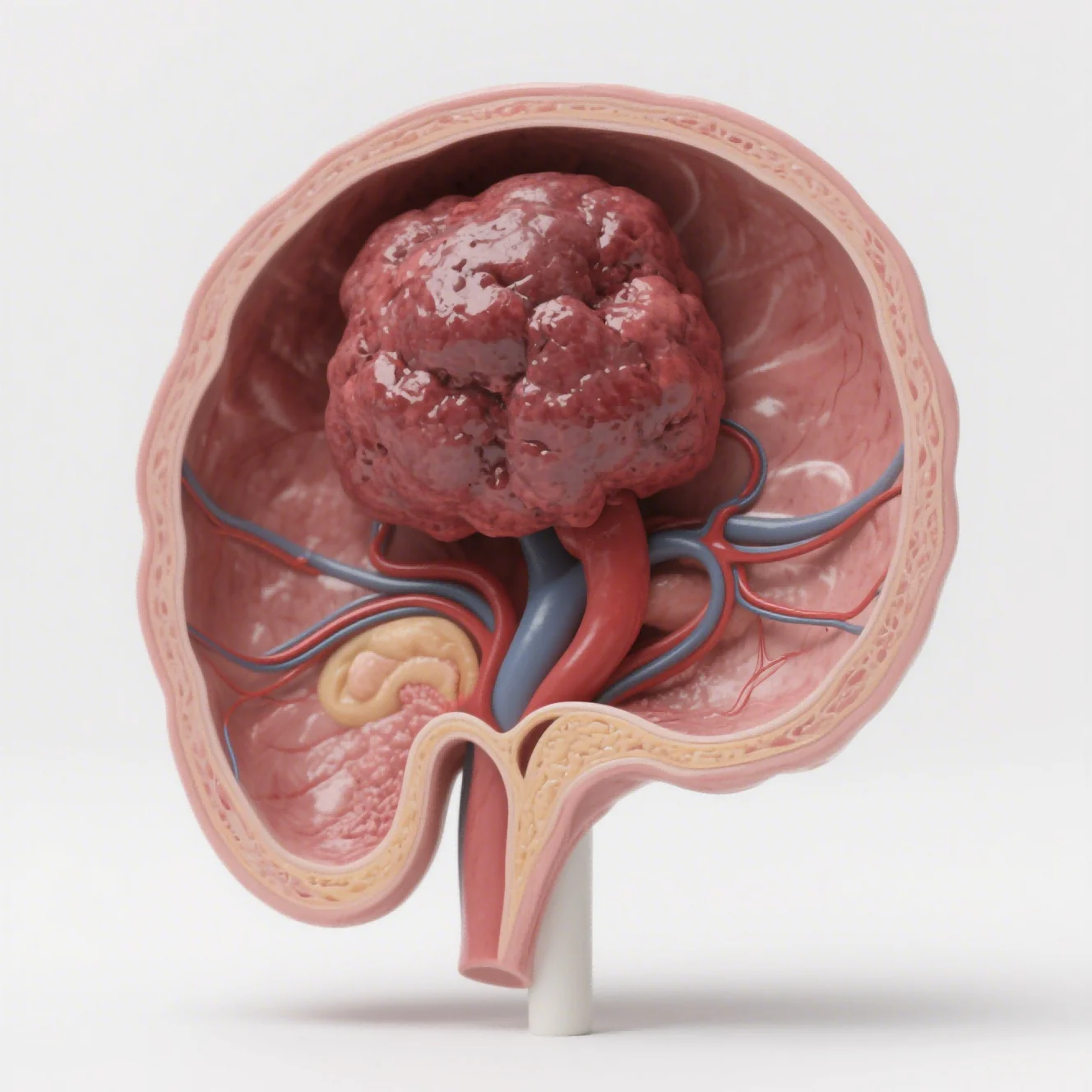
Recreate Tumor Growth in the Relevant Organ
We support fluorescence or bioluminescence imaging, ultrasound, H&E, IHC, flow cytometry, qPCR, and ELISA as supporting technologies for orthotopic research. We match each orthotopic pancreatic study method to the evidence the program needs. For each orthotopic option, the study team examines how faithfully the selected model reflects the relevant tumor biology and treatment mechanism.
Practical efficiency is useful only after the orthotopic pancreatic study design remains scientifically fit. We establish the pancreatic-model record with source material, implantation details, baseline burden, and imaging criteria. The orthotopic pancreatic study team agrees on the study model, checks, and acceptance criteria together.
We explain that conventional subcutaneous grafts are convenient and widely used, but they do not reproduce the organ-specific microenvironment that influences tumor-host interaction. The study plan addresses angiogenesis, invasion, metastasis, drug distribution, metabolism, and treatment response.
Our orthotopic or in situ platform implants tumor cells or patient-derived tissue into the anatomically corresponding organ of immunodeficient mice, including liver, stomach, cervix, and other sites. For the evidence question at hand, we make the biological reach and omissions of the orthotopic pancreatic model explicit.
Once dosing or measurement starts, organ-specific growth, dissemination, and tissue response guide the next pancreatic-model comparison. The study team chooses the pancreatic model around organ-specific growth, dissemination, and the intended treatment mechanism. Our applications include orthotopic liver, gastric, and cervical cancer, lung and bone metastasis, integrated primary-and-metastatic models.
For broader orthotopic pancreatic study questions, we consider orthotopic PDX models and, where appropriate, humanized immune-system models for immunotherapy evaluation. We select the orthotopic pancreatic study system for the specific decision rather than for universal convenience. In Jennio Biotech orthotopic studies, we keep the purpose of each technical choice visible.
Monitor a Lesion That Cannot Be Measured by Calipers
Technical readouts include fluorescence or bioluminescence imaging, small-animal ultrasound, H&E, IHC, flow-cytometric immune profiling, qPCR, ELISA, body weight, survival, tumor weight, and pathology. This phase evaluates measurement consistency and model performance rather than relying on visual appearance alone
Our program team reviews variation, deviations, and assay performance as data accrue. As the orthotopic pancreatic study protocol runs, we can now compare alternative models by the functions they add and the limitations they introduce. Openness about methods and deviations strengthens experimental review.
Our process includes requirement confirmation, cell-line or PDX-source selection, endpoint and dosing alignment, and model establishment and validation, which typically require two to four weeks depending on the selected model and study design. Our orthotopic pancreatic study log captures exclusions and unexpected outcomes in real time.
The study team retains implantation, imaging, pathology, and dosing records so outside reviewers can reconstruct the orthotopic pancreatic reasoning. When interpreting the result, we include three to eight weeks of efficacy work, and subsequent data analysis with raw images, statistics, IHC images, and a GLP-aligned report.
Here, chronology is part of the biology. The pancreatic model schedule accounts for surgery, engraftment, imaging, dosing, metastatic assessment, and tissue collection. The central inquiry shifts from system choice to measurement discipline. We do not aggregate study observations in ways that obscure animal-level or sample-level data.
Contemporaneous notes preserve the boundary between the orthotopic pancreatic study plan and subsequent exceptions. The current judgment can change when a different model or method adds material information. We use that boundary to keep interpretation proportionate to the orthotopic pancreatic study results.
Control Surgical Variability and Interpretation
Quality controls include STR authentication, pathogen testing, SOP management, double-blind imaging review, cross-review of tissue sections, electronic records, and multilevel review before delivery. We bring separate results together when judgment reaches its decision point. We consider the orthotopic pancreatic study response alongside exposure, tissue findings, mechanism, and safety signals.
Our operations maintain an SPF animal center with in vivo imaging, small-animal ultrasound, and microsurgical support. We keep the pancreatic model’s limits visible in the final interpretation. Our pancreatic-model report states which organ-specific finding was observed and which patient-level question remains unanswered.
Our pancreatic orthotopic system workflow links imaging, pathology, survival, and metastatic assessment. The study team chooses the model by the study question: subcutaneous systems support efficient screening and caliper measurement. Orthotopic systems instead address local growth, organ physiology, invasion, metastasis, and tissue-specific exposure.
In practical execution, traceability strengthens the reliability of the final interpretation. We preserve a documented chain linking orthotopic pancreatic study protocol choices to deviations, analysis, and source data. Our final step turns the findings into an explicit choice.
Neither format removes the need for appropriate controls, validated implantation, prospective success criteria, serial monitoring, ethical oversight, and careful interpretation of model limitations. Our final evaluation records why the initiative advances or changes direction. The Jennio Biotech team presents the pancreatic-model finding alongside its raw basis and constraints.