
Cancer biology changes with location. A malignant cell growing beneath the skin encounters a very different environment from one developing inside the organ where the disease normally arises. An orthotopic model of tumor places cancer cells or tissue into the corresponding organ or tissue of origin, allowing researchers to examine growth, invasion, local interactions, and treatment response under conditions that more closely resemble selected aspects of the original disease.
We at Jennio Biotech use this type of model as one part of a broader preclinical strategy, with study design determined by the biological question rather than by model convenience alone.
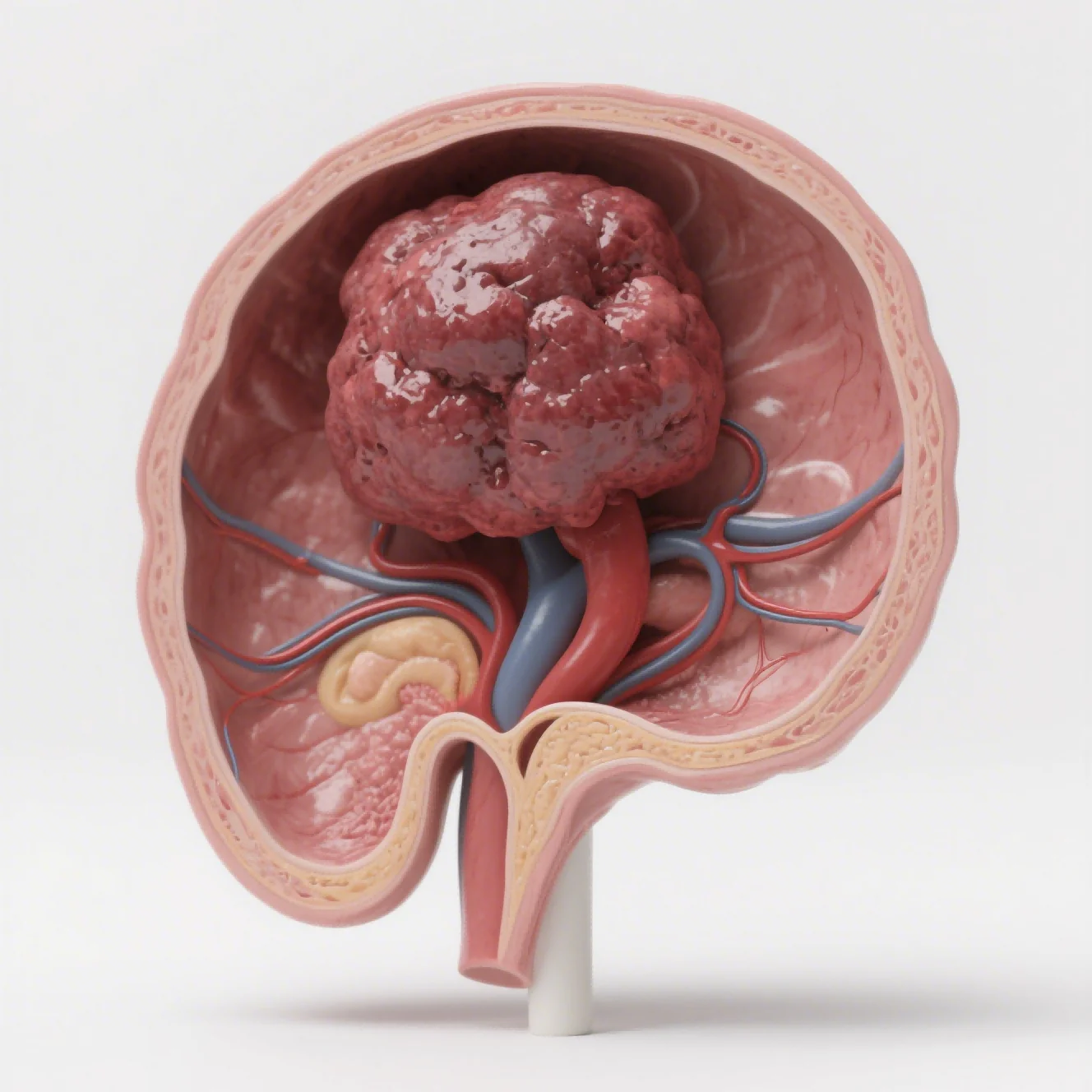
Recreating the Native Tumor Environment
Tumor cells interact continuously with neighboring tissue, extracellular matrix, blood vessels, and stromal components. Their anatomical surroundings can influence proliferation, vascular development, invasion, and response to treatment, making location an important variable in experimental cancer research.
A conventional subcutaneous xenograft is useful for many efficacy studies because tumor growth can be monitored relatively easily. Its ectopic location, however, may not reproduce tissue-specific signals or local physiological conditions associated with the original malignancy.
Placing tumor cells within the corresponding organ changes the experimental context. Liver, gastric, and cervical tumors, for example, can develop within tissues that provide distinct cellular and vascular environments. Such differences may influence both tumor progression and the way a compound reaches its intended target.
Exploring Growth, Invasion, and Metastasis
Anatomical placement can provide information beyond simple tumor enlargement. Researchers may investigate whether malignant cells invade surrounding structures, alter local tissue organization, or develop characteristics associated with disease progression.
Imaging becomes particularly valuable when tumors are difficult to monitor through external measurements. With appropriate fluorescent or luciferase reporters, fluorescence or bioluminescence imaging can allow repeated observation of tumor-associated signals over time, helping researchers compare disease trajectories between treatment groups without relying exclusively on endpoint measurements.
Metastatic behavior requires another level of consideration. A primary tumor model may describe local progression, whereas dedicated metastatic systems can investigate dissemination to organs such as the lung or bone. The appropriate choice depends on whether the study is examining primary tumor control, metastatic spread, or both.
Connecting Models with Therapeutic Questions
Drug response is shaped by more than the intrinsic sensitivity of cancer cells. Local vascularization, tissue penetration, cellular interactions, and disease architecture can all affect pharmacological activity. An organ-specific model can provide additional evidence when these factors are central to the mechanism being investigated.
Our model applications at Jennio Biotech include orthotopic liver cancer using intrahepatic implantation of SMMC7721 luc cells, cervical cancer using cervical transplantation of HeLa luc cells, and gastric cancer through gastric-wall implantation of MKN45 luc cells. In representative studies, cisplatin reduced tumor volume in the cervical model, while oxaliplatin reduced tumor-associated bioluminescence and tumor weight in the gastric model.. These examples illustrate how imaging and endpoint measurements can be combined to assess treatment response.
Different therapeutic categories may require different evidence. A cytotoxic compound may be evaluated primarily through tumor burden and tissue morphology, while a targeted agent could require molecular markers. Immunotherapy studies introduce additional considerations because immune-cell composition and cytokine activity become relevant experimental variables.
Adding Imaging and Tissue-Level Evidence
Bioluminescence and fluorescence imaging can provide longitudinal information that a single endpoint cannot capture. Changes in signal intensity may help researchers follow disease progression or treatment-associated changes across multiple time points, but should be interpreted alongside reporter expression, tissue attenuation, and other factors affecting signal detection.
Histopathology adds structural evidence. H&E staining can reveal tissue architecture and pathological changes, while immunohistochemistry can examine selected proteins or cellular markers. These methods can help connect macroscopic treatment effects with changes occurring inside the tumor.
Flow cytometry and molecular assays further expand the analytical framework. Immune-cell phenotyping can characterize components of the tumor microenvironment, while qPCR or ELISA may be used to examine gene expression or cytokine-related responses. Combining complementary measurements can make biological interpretation more informative than relying on tumor size alone.
Extending Research to Immune and Metastatic Settings
Some research questions cannot be addressed adequately through a primary tumor system alone. Metastatic models can focus on dissemination, while immune-oriented designs examine interactions between treatment and host immune components.
Tail-vein administration of Nalm 6 leukemia cells, for instance, can be used to investigate leukemia-associated dissemination and treatment response through imaging. Intra-tibial implantation of breast cancer cells provides another experimental route for studying tumor growth within bone and treatment effects on tumor-associated bone destruction, but does not reproduce the earlier steps of metastasis to bone.
Humanized immune-system models offer another dimension. Human PBMCs or CD34+ stem cells can be introduced into NCG mice before orthotopic tumor establishment, creating a framework for evaluating agents such as PD-1 or CTLA-4 antibodies alongside tumor-infiltrating lymphocytes and cytokine secretion. Such designs are particularly useful when immune mechanisms form a central part of the research hypothesis.
Conclusion
Cancer models become more informative when their biological context matches the question under investigation. An orthotopic model of tumor can better reflect organ-specific anatomical relationships than an ectopic model, while imaging, pathology, flow cytometry, and molecular testing can provide complementary evidence of tumor growth, tissue changes, and relevant cellular or molecular responses.
Our work at Jennio Biotech combines these approaches according to tumor type, therapeutic mechanism, and research stage, allowing investigators to examine efficacy alongside invasion, metastasis, tissue response, and immune activity. The value of an orthotopic system ultimately comes from using its biological context thoughtfully rather than treating it as a universal substitute for every other model.