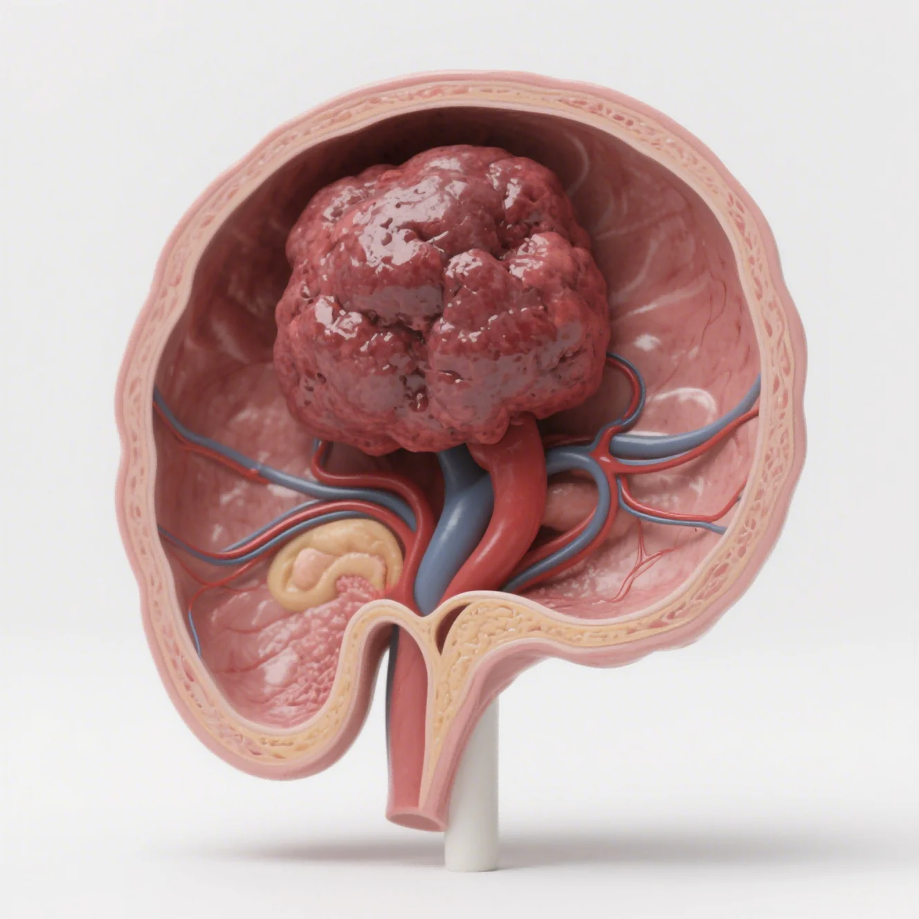
Inflammation and immunology disease models become informative when induction, pathway biology, and endpoints represent relevant aspects of human disease biology. We then connect in-life disease scores, immune phenotyping, cytokines, and pathology in one mechanistic evidence record. We therefore begin with the uncertainty that the supporting information needs to reduce.
Our capabilities include preclinical models such as CIA or AIA for arthritis, DSS or TNBS for colitis, imiquimod for psoriasis-like skin disease, EAE for neuroinflammation, and additional autoimmune or allergic models, subject to project-specific model qualification. This distinction matters because experimental system fit and execution quality influence every later interpretation.
The documented induction method, pathway, and endpoint panel show what another inflammation model can contribute. For this inflammation and immunology question, missing raw observations or unexplained deviations weaken the link between a reported value and the underlying biology. Applications include screening anti-inflammatory agents, immunosuppressants, antibodies, JAK or cytokine-pathway inhibitors, and other biologics.
We limit the inflammation and immunology study claim to the evidence collected. Clinical scores and body weight show whole-animal status, while cytokine profiling, Th17/Treg analysis, pathology, IHC, and immunofluorescence reveal mechanism and tissue change.
Humanized may support the evaluation of human-specific antibodies when the model provides relevant target expression or immune-cell reconstitution. For us, useful evidence combines biological relevance, transparent operations, and a disciplined next-step determination. We explain to our partner how the inflammation and immunology study evidence supports the interpretation.
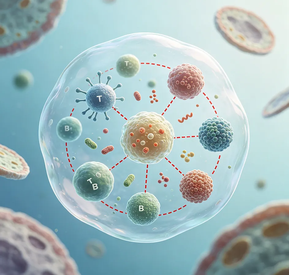
Select Models by Pathology and Therapeutic Target
Our first basis for interpreting an inflammation and immunology study is appropriately characterized and suitable biological material. Viewed as one inflammation and immunology dataset, our checks cover inflammation and immunology study provenance, identity, condition, and suitability. A useful review needs to explain both the translational value and the limitations caused by species differences, induction variability, and incomplete reproduction of chronic human disease.
While we review inflammation and immunology, our inflammatory and autoimmune platform includes preclinical models relevant to rheumatoid arthritis, inflammatory bowel disease. The same evaluation considers psoriasis, multiple sclerosis, lupus, gout, allergic disease, and other immune-mediated conditions. Taken together in inflammation and immunology, we use an assay for inflammation and immunology study only when it represents the relevant biology.
For inflammation and immunology studies, we record the system, dose, timing, and readout boundary before interpretation. The study team judges inflammation and immunology study against the research question rather than the convenience of the setup. A standard service description still requires proof that the inflammation and immunology study system performs as intended.
Across the project, our work explains that inflammation is a protective immune response to injury or infection but may become pathogenic when activation is prolonged or excessive. We examine the results supporting the biological model or assay itself.
We organize inflammation & immunological disease models by pathology, therapeutic target, and disease stage. Available study models include collagen-induced or adjuvant-induced arthritis, DSS- or TNBS-induced colitis, imiquimod-induced psoriasis-like skin inflammation, EAE, and other validated mouse or rat systems.
In practice, before discussing throughput, we establish relevance. The development effort gains value when the biological model mirrors the mechanism under review. Our delivery includes the scientific reasoning behind the inflammation and immunology study conclusion.
Combine Clinical Scores with Mechanistic Endpoints
For inflammation and immunology studies, the measurement strategy defines which in-life, cellular, and tissue-level questions we can answer. As the inflammation and immunology plan develops, the evaluation framework combines body weight, lesion or disease scores, swelling, behavioral measures, organ indices, survival or recurrence.
We keep Luminex cytokine profiling, flow-cytometric immune phenotyping such as the Th17/Treg balance, H&E, Masson staining, IHC, immunofluorescence, and digital pathology traceable. Together, these readouts connect changes in disease phenotype with cellular and tissue mechanisms.
When the inflammation and immunology evidence must guide action, operational consistency protects scientific interpretation. Across the complete record, our study plan coordinates sample timing with equipment and reviewer availability.
Our inflammation & immunological disease models combine clinical observations with mechanistic and tissue-level endpoints. Execution records allow the final inflammation and immunology study figures to remain traceable. Our records link each observation to its method, material, and check status.
For psoriasis, our workflow specifically mentions PASI-like scoring, epidermal thickness, skin pathology, and inflammatory mediators such as IL-17, IL-23, IL-12, and TNF-alpha. For Jennio Biotech inflammation studies, we keep the reasoning, execution record, and limitations open to review.
At the related stage, we report a library of more than 50 validated inflammatory or autoimmune models, multicolor flow cytometry. The handoff makes high-throughput cytokine testing, digital pathology, and humanized immune-system mice for human-specific antibodies reviewable.
Early discussion of small execution inconsistencies protects the intended immunology comparison. In practice, this safeguard reduces inflammation and immunology study rework and protects the intended comparison. Progression of the inflammation and immunology study program is tied to stage-specific findings.
Translate Carefully Across Species and Disease Stages
Our immunology closeout joins in-life, cellular, soluble-marker, and tissue-level evidence while documenting discordant findings for review. We trace the inflammation and immunology study conclusion from predefined thresholds to individual observations. At that stage, the workflow covers alignment on the model, groups, dosing, and endpoints; induction and administration; dynamic sharing of body weight.
The program can draw on behavior, and other progress data; sample collection; molecular and pathological analysis; and a GLP-aligned final package with raw data and statistics. At the action threshold, a useful conclusion includes both the finding and its unresolved limits. Our final package identifies both the supported conclusion and the unresolved objective.
The handoff passes only when the inflammation and immunology study reasoning is understandable without the original study team. At this inflammation and immunology step, quality statements include AAALAC animal-welfare guidance, ISO 9001 quality management, electronic laboratory notebooks. The same evaluation also covers negative and positive controls, independent review of key measurements, and traceable records.
The delivered inflammation and immunology study package carries study logic and limits with the numerical results. We review model relevance, induction consistency, controls, scoring, and blinding with the partner before execution. The study team connects sample handling, calibration, exclusions, statistics, welfare safeguards, raw-data access, and deviations to the main finding.
The final point turns observations into a bounded development action threshold. When the inflammation and immunology findings are combined, we separate what the research plan demonstrates from what still requires confirmation. Our Jennio Biotech interpretation cross-checks clinical signs, immune markers, cytokines, and pathology.